Keyhole removal of gallstones and the gallbladder for stones, chronic cholecystitis and polyps, minimal pain, 1-day hospital stay, back to normal life in 3–5 days.


Gallstones at a Glance
The gallbladder is a small, pear-shaped organ tucked beneath the liver that stores bile, a digestive fluid produced by the liver to break down dietary fats. Bile contains cholesterol, bile salts, and bilirubin. When these components fall out of balance due to diet, genetics, obesity, or hormonal changes, they crystallise and form gallstones. These range from a grain of sand to a golf ball, and most people develop multiple stones simultaneously. Cholesterol stones (80%) are the most common type.
Once gallstones become symptomatic, the risk of serious complications rises significantly. Acute cholecystitis (gallbladder inflammation), choledocholithiasis (stones blocking the bile duct), and pancreatitis can all develop, each requiring increasingly complex emergency management. Elective laparoscopic cholecystectomy before complications arise is far safer, faster, and less disruptive than emergency surgery. The procedure eliminates future gallstone risk entirely.
Reassuringly, the gallbladder is not essential to life. The liver continues producing bile, which drips directly into the small intestine. The vast majority of patients adapt within weeks and resume a completely normal diet and lifestyle.
Book a consultation with Dr. Prashanth J V today.
Book Consultation 📞 +91 93533 16175The entire procedure is performed under general anaesthesia and takes approximately 45–60 minutes. You are completely comfortable throughout.
You are comfortably put to sleep. The abdomen is cleaned and monitoring lines are placed. The anaesthesiology team ensures your complete comfort throughout.
3–4 tiny incisions (5–10 mm each) are made. Carbon dioxide is gently introduced to inflate the abdomen, creating a safe working space between the organs.
A high-definition laparoscope is inserted, projecting real-time visuals on a monitor. Dr. Prashanth can see the gallbladder and surrounding structures with exceptional clarity.
Specialised instruments clip and divide the cystic duct and cystic artery. The gallbladder is carefully freed from the liver bed. Critical view of safety is confirmed to protect the common bile duct.
The gallbladder (containing all stones) is placed in a retrieval bag and removed through one port. Incisions are closed with absorbable sutures or skin glue, no stitches to remove.
Rest at home. Mild soreness around incision sites managed with regular analgesics. Discharged the morning after surgery in most cases.
Walking freely around the house. Soft diet progresses to normal food. Most shoulder-tip gas pain (from CO2) resolves.
Return to desk work. Avoid heavy lifting and strenuous exercise. Driving resumes once off strong pain medication.
Full unrestricted activity including exercise and heavy lifting. A small minority may notice temporary loose stools after very fatty meals, which resolves as the body adapts.
Gallstones exist on a spectrum from silent stones causing nothing at all, through to life-threatening infection. Recognising which stage you are at determines how urgently something needs to be done.
| Stage | What happens | What it means |
|---|---|---|
| Silent stones | Stones present but causing no symptoms. Often found incidentally on a scan done for another reason. | Usually no treatment needed. A minority go on to develop symptoms each year. |
| Biliary colic | A stone temporarily blocks the gallbladder outlet. Severe upper abdominal pain lasting from thirty minutes to a few hours, then settling. | The gallbladder is not infected. Surgery is generally advised, as attacks recur. |
| Acute cholecystitis | The blockage persists and the gallbladder becomes inflamed and infected. Constant pain, tenderness and fever. | Needs admission, antibiotics and usually surgery, often during the same admission. |
| Choledocholithiasis | A stone escapes into the bile duct and obstructs it. Jaundice, dark urine, pale stools and itching. | The duct stone must be cleared, usually by ERCP, before or alongside gallbladder removal. |
| Cholangitis | Infection of the obstructed bile duct. Pain, jaundice and high fever with rigors. | A medical emergency requiring urgent admission, antibiotics and drainage of the duct. |
| Gallstone pancreatitis | A stone obstructs the pancreatic duct outlet, inflaming the pancreas. Severe upper abdominal pain radiating to the back, with vomiting. | Requires urgent hospital admission. Gallbladder removal follows to prevent a further attack. |
The important practical point is that biliary colic is a warning rather than a nuisance. Attacks tend to recur and become more frequent, and a proportion of people go on to develop one of the complications above. Planned surgery in a calm setting is considerably safer than emergency surgery during acute inflammation.
Bile contains cholesterol, bile salts and pigments in a delicate balance. When it holds more cholesterol than the bile salts can keep dissolved, crystals form, and those crystals aggregate into stones. Sluggish emptying of the gallbladder gives them time to grow.
Oestrogen increases cholesterol secretion into bile and progesterone slows gallbladder emptying.
Prevalence rises steadily with each decade of life.
Hormonal changes and reduced gallbladder emptying make stone formation more likely.
Associated with higher cholesterol saturation of bile.
Including after bariatric surgery or crash dieting, which markedly increases stone formation.
Associated with altered bile composition and impaired gallbladder motility.
There is a clear inherited component to stone formation.
Conditions such as thalassaemia and sickle cell disease produce pigment stones from excess bilirubin.
Bile stagnates in a gallbladder that is not being emptied regularly.
Including some lipid-lowering drugs and oestrogen-containing preparations.
The traditional teaching of the four Fs, female, forty, fertile and overweight, captures several of these but is an oversimplification. Gallstones occur in men, in the young, and in people of normal weight.
Diagnosis combines a characteristic history with imaging, and additional tests are added where a complication is suspected.
History and examination. The pattern of pain is often diagnostic on its own. Severe, constant pain in the upper right abdomen or epigastrium, radiating to the back or right shoulder, coming on after a meal and lasting hours. Tenderness under the right ribs that catches the breath on deep inspiration suggests inflammation of the gallbladder.
Ultrasound of the abdomen. The first-line investigation and highly reliable for detecting stones in the gallbladder. It also shows thickening of the gallbladder wall, fluid around it and dilatation of the bile ducts, all of which point to complications. It is quick, painless and involves no radiation.
Blood tests. Liver function tests, full blood count and amylase or lipase. Raised bilirubin and alkaline phosphatase suggest a stone in the bile duct. A raised amylase or lipase suggests pancreatitis. These results frequently change the plan.
MRCP. A specialised MRI of the biliary tree, used when a duct stone is suspected on blood tests or ultrasound. It maps the ducts without instrumentation and determines whether ERCP is needed.
Why this sequence matters. Missing a stone in the bile duct and proceeding straight to gallbladder removal leaves the duct stone behind, which can then cause jaundice or pancreatitis afterwards. Establishing the state of the ducts before surgery avoids this.
The decision rests on whether the stones are causing symptoms, and on whether any complication has occurred.
| Surgery usually advised | Usually managed without surgery |
|---|---|
| Recurrent biliary colic affecting daily life | Silent stones found incidentally, with no symptoms at all |
| Acute cholecystitis, ideally during the same admission | Vague indigestion or bloating with stones present, where the stones may be coincidental |
| A previous episode of gallstone pancreatitis | Patients unfit for anaesthesia, where symptoms are controlled medically |
| Stones with a bile duct stone, after the duct has been cleared | Pregnancy in the first and third trimesters, where surgery is generally deferred if possible |
| A large stone burden, gallbladder polyps or a calcified gallbladder wall | Asymptomatic stones in the very elderly with significant comorbidity |
There is one important qualification about silent stones. A calcified, so called porcelain gallbladder, or gallbladder polyps above a certain size, are associated with a higher risk of gallbladder cancer and removal is advised even without symptoms. Gallbladder cancer is uncommon, but its incidence in parts of India is higher than in many other countries, which makes this a relevant consideration rather than a theoretical one.
Diagnosed with gallstones?
Not every gallstone needs surgery. A consultation reviews your ultrasound and symptoms and tells you whether removal is advisable, and when. Severe pain with fever or yellow eyes needs same-day care.
Preparation for a planned laparoscopic cholecystectomy is routine.
Laparoscopic cholecystectomy is among the most commonly performed abdominal operations and is generally very safe. One specific complication, injury to the bile duct, is rare but serious enough that it deserves to be named explicitly rather than buried in a list.
| Effect | How often | What it means |
|---|---|---|
| Shoulder tip pain | Common | Caused by gas used to inflate the abdomen irritating the diaphragm. Settles within a day or two. |
| Wound soreness and bruising | Common | At the four small port sites. Eases over one to two weeks. |
| Nausea | Common | Related to anaesthesia, and usually short-lived. |
| Looser stools after fatty meals | Occasional | Bile now enters the intestine continuously. Usually settles within weeks to months as the body adapts. |
| Conversion to open surgery | Uncommon | Where inflammation or scarring makes the anatomy unsafe to define. This is a judgement in favour of safety, not a complication. |
| Bleeding or wound infection | Uncommon | Usually manageable without further surgery. |
| Retained bile duct stone | Uncommon | A stone left in the duct, causing jaundice or pain afterwards. Treated by ERCP. |
| Bile leak | Uncommon | Bile escaping from the gallbladder bed or a small duct. May need drainage or ERCP. |
| Bile duct injury | Rare but serious | Damage to the main bile duct, which can require major reconstructive surgery. Prevented by careful dissection and confirming the anatomy before dividing anything. |
| Port site hernia | Rare | A hernia through one of the small incisions, which may need repair. |
For symptomatic gallstones, surgical removal of the gallbladder is the definitive treatment. The alternatives are limited and are reserved for specific situations.
| Treatment | How it works | Durability | Best suited to |
|---|---|---|---|
| Observation | No intervention. Symptoms and complications are monitored. | Not applicable | Silent stones causing no symptoms. |
| Dietary modification | Reducing fatty foods may reduce the frequency of attacks. | Controls symptoms only | Temporary measure while awaiting surgery. |
| Laparoscopic cholecystectomy | The gallbladder is removed through four small incisions. The standard operation. | Definitive | Almost all symptomatic gallstones. |
| Open cholecystectomy | Removal through a single larger incision under the ribs. | Definitive | Severe inflammation, difficult anatomy, or conversion from laparoscopic. |
| ERCP | An endoscope is passed to the bile duct opening and stones are removed from the duct itself. | Clears the duct only | Bile duct stones, usually before or after gallbladder removal. |
| Percutaneous cholecystostomy | A drain is placed into the gallbladder through the skin to control infection. | Temporary | Very unwell patients unfit for immediate surgery. |
| Bile acid dissolution therapy | Oral medication to slowly dissolve small cholesterol stones. | Slow and often incomplete, with high recurrence | Rarely used. Occasionally where surgery is not possible. |
Shock wave lithotripsy, which is used for kidney stones, is not an effective treatment for gallstones. The fragments remain in a diseased gallbladder and stones reform.
Silent stones can reasonably be left alone. Stones that have caused an attack of biliary colic are a different matter, because that first attack is usually not the last.
Attacks recur and intensify. Most people who have had one episode of biliary colic go on to have more, often with increasing frequency, and the interval between them tends to shorten.
Progression to acute cholecystitis. Persistent obstruction leads to infection of the gallbladder, requiring hospital admission and antibiotics. Surgery in this setting is technically more difficult and carries a higher chance of conversion to open surgery.
Stones escaping into the bile duct. This produces jaundice, and if the duct becomes infected, cholangitis, which is a life-threatening emergency.
Gallstone pancreatitis. One of the most serious complications, in which a stone obstructs the pancreatic duct outlet. Severe pancreatitis can be life-threatening and requires intensive treatment.
The core argument. A planned operation in a calm, prepared setting is considerably safer than the same operation performed as an emergency during acute inflammation. That is the main reason surgery is advised after symptoms begin rather than waiting for a complication to force the issue.
The gallbladder concentrates and stores bile between meals. Without it, the liver produces bile continuously and it drains straight into the intestine. Digestion adapts, and the vast majority of people eat completely normally.
Removing the gallbladder does not increase the risk of any long-term illness in a way that should influence the decision, and it does not require lifelong medication or dietary restriction.
#38/2, 2nd Main Road, Madiwala New Extension, Bangalore 560068, behind Ayyappa Temple. Convenient for BTM Layout, Koramangala, HSR Layout, Bommanahalli and Hosur Road.
Dr. Prashanth J V also consults in Koramangala, Jayanagar and Seshadripuram. When you call, the team will confirm the most convenient location for gallbladder stones.
Consultations are by appointment. Call +91 93533 16175 or request an appointment online.
Laparoscopic cholecystectomy has been a core part of his practice for over two decades, performed with the critical view of safety on every case.
Latest HD equipment, advanced energy devices, and meticulous critical view of safety to protect the common bile duct.
Most patients go home the morning after surgery, minimising hospital stay and dramatically reducing hospital-acquired infection risk.
Medical References & Further Reading
This page is written and medically reviewed by Dr. Prashanth J V, MBBS, MS, FMAS, FISCP, DMAS, Karnataka Medical Council Registration No. 41048. It is general health information about gallstone disease and does not replace an individual consultation, examination and investigation. If you have symptoms, please seek personalised medical advice.
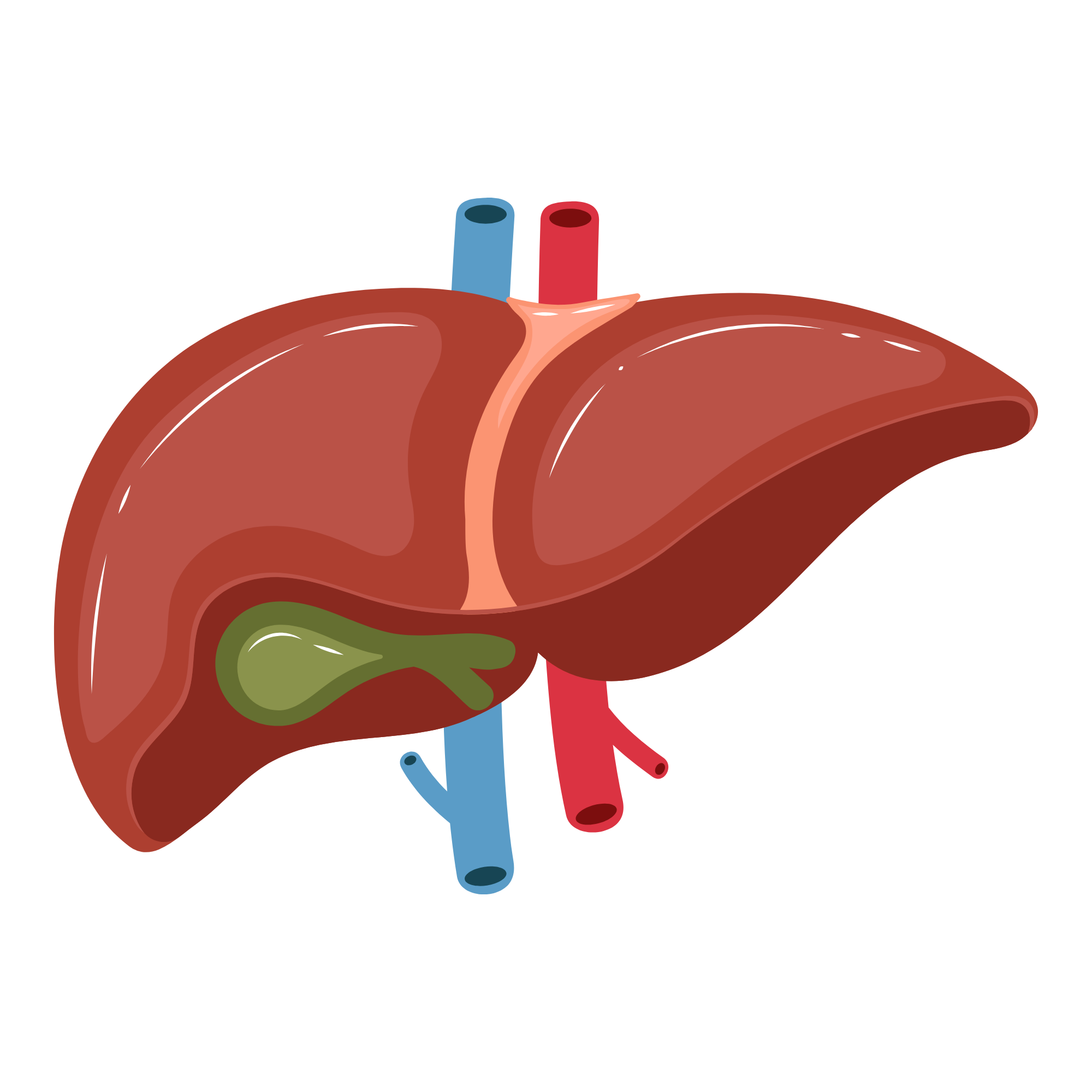 Gallbladder Surgery
 Appendicectomy
 Hernia Repair
 Hysterectomy
 Ovarian Cystectomy
 Tubectomy & Ectopic
 Laser Piles
 Laser Fissure
 Varicose Veins
 Laser Fistula
 Pilonidal Sinus